
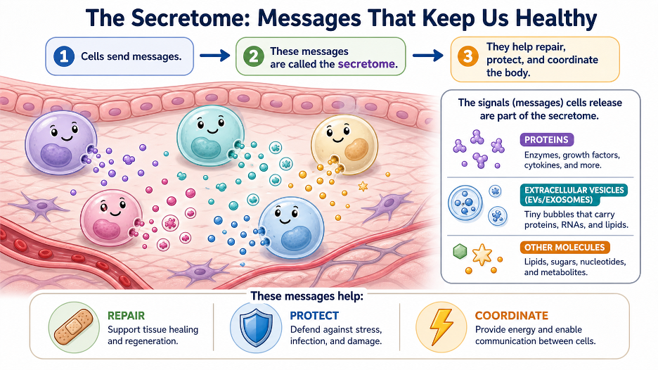
Over the last decade, scientists have realized that most of the benefit from stem cell therapies does not come from the cells turning into new tissue, but from the molecules they release into their surroundings. These secreted molecules — the secretome — may ultimately be more useful than giving patients whole stem cells or isolated exosomes alone. The challenge now is not whether the secretome works, but how to standardize, scale, and integrate it into existing regulatory pathways. I am excited about this new modality and its potential.
What the Secretome Actually Is
The secretome is not just exosomes. It is the complete collection of biologically active substances that stem cells release into the surrounding fluid. This includes:
- Extracellular vesicles (EVs), such as exosomes and microvesicles, carry mRNA, microRNA, and proteins between cells.
- Soluble growth factors, such as VEGF, HGF, IGF, FGF, TGF‑β, and HIF, which drive new blood vessel growth, cell survival, and tissue repair.
- Cytokines and chemokines, like CCL2, CCL5, CCL7, and TNF‑β, which help dial the immune system up or down.
Non-coding RNAs, including circular RNAs and long non‑coding RNAs, can reprogram how recipient cells read their genes.
Exosomes are therefore one piece of a much larger communication toolkit heavily studied and commercially emphasized, but far from the whole story.
Why the Secretome May Outperform Exosomes Alone
The Synergy Argument
Mesenchymal stromal cell (MSC) therapies now appear to work primarily through a coordinated mix of secreted factors, rather than a single type of vesicle or molecule. Exosomes are powerful messengers, but they represent only one channel in that conversation. The full secretome simultaneously delivers:
- Anti‑inflammatory cytokines that shift immune cells (like macrophages) into a more healing, pro‑repair state.
- Angiogenic factors that promote blood vessel remodeling and restore circulation.
- Anti‑apoptotic signals that prevent stressed or injured cells from dying prematurely.
- EVs encapsulated microRNAs that reprogram gene expression inside target cells.
When you isolate only exosomes and discard the rest, you lose much of this orchestrated, multi‑layered signaling environment. Studies comparing adipose-derived small EVs with full conditioned medium show that each has distinct biological signatures, and neither clearly wins in all situations, reinforcing that context and synergy matter.
Secretome vs. Live Stem Cells: Practical and Safety Benefits
Compared with infusing live stem cells, using the secretome (including its exosomes) offers several attractive advantages:
- Far lower tumor risk, because no dividing cells are being implanted.
- Minimal immune rejection, since the product is mostly signaling molecules rather than whole foreign cells.
- Improved storage: secretome preparations can be frozen (for example, at −80 °C) and kept stable for long periods.
- Multiple doses from a single cell harvest are especially useful for chronic diseases requiring repeated treatment.
- Less manufacturing complexity than exosome‑only products, which require additional isolation and purification steps.
- A likely clearer long‑term regulatory path as a biologic or drug, once standards for production and testing mature.
In simple terms, the secretome retains the benefits of stem cell-derived signals while eliminating most of the logistical and safety issues associated with live cells.
Preconditioning: “Training” Cells to Produce a Better Secretome
One of the most compelling aspects of secretome therapy is that you can shape what cells secrete by how you culture them before collection. Preconditioning strategies include:
- Hypoxia (low oxygen, such as 2%): mimics an ischemic or low-oxygen injury environment, prompting cells to release more neuroprotective, blood vessel-building, and anti-inflammatory factors.
- Inflammatory priming (for example, IFN-γ, TNF-α) boosts the immunomodulatory, “immune calming” component of the secretome.
- Three‑dimensional culture (spheroids, bioreactors): enhances EV output and concentrates the secretome.
- Serum‑free or Xeno‑free conditions: reduce animal-derived contaminants and help meet clinical-grade manufacturing standards.
This tunability means that, in principle, you can design different secretomes for orthopedics, neurology, cardiometabolic disease, or oncology, instead of relying on a “one‑size‑fits‑all” exosome product.
The Case Against Exosomes as a Standalone Platform
Exosomes remain a promising tool, but several practical issues have slowed their path as a stand‑alone solution:
- Isolation loss: Common isolation methods (such as ultracentrifugation or size-exclusion chromatography) can lose a large fraction of vesicles and may entrain unwanted contaminants.
- Batch variability: Without strict, harmonized protocols, exosome batches can differ widely in cargo, surface markers, and potency, even when derived from similar cells.
- Incomplete picture: Purified exosome products leave behind the soluble growth factor and cytokine fraction, which is rich in VEGF, HGF, and other regenerative molecules.
- Regulatory ambiguity: There is still no fully mature, harmonized regulatory pathway for exosome products, complicating clinical approval and reimbursement.
These challenges do not render exosomes useless. Instead, they underscore why the full secretome, which preserves all components and avoids additional isolation steps, may be more efficient and reliable in day-to-day clinical practice.
Where the Secretome Still Struggles
Despite its promise, secretome therapy has its own obstacles:
- Lack of standardization: There is no universal agreement yet on how to grow cells, when to collect the secretome, how to concentrate it, or which potency tests best predict clinical outcomes.
- Scalability and cost: Producing large volumes under full GMP conditions is technically demanding and expensive.
- Regulatory gray zone: The secretome does not fit neatly into traditional drug or biologic categories in many jurisdictions, which slows approvals.
- Variable composition: The secretome profile varies with donor age, cell type, passage number, and culture conditions, introducing noise and variability.
Tumor biology considerations: In some situations, specific secretome profiles may promote tumor growth or spread, so oncology applications require careful design and screening.
How Secretome, Exosomes, and Stem Cells Fit Together
Rather than viewing these options as competitors, it is more useful to think of them as complementary tools, each suited to particular jobs:
- Stem cells: Reserved for situations where long‑term engraftment, tissue replacement, or true hematopoietic reconstitution are needed.
- Isolated exosomes: Used when nanoscale vesicles offer unique benefits, such as crossing certain biological barriers (for example, into the central nervous system) or delivering drugs and genetic cargo.
- Full secretome: Used when broad, systemic anti‑inflammatory and pro‑regenerative effects are desired, especially in chronic conditions needing repeated dosing.
Clinical trials are already exploring MSC‑derived secretome in osteoarthritis, chronic pain, neurologic repair, skin disease, and other areas, with early evidence accumulating but still incomplete.
MUSE Cells: A Next‑Level Secretome
If the MSC secretome is a step beyond exosomes alone, then the secretome from MUSE cells is another jump beyond that. MUSE (Multilineage-differentiating Stress-Enduring) cells are a small, specialized subset of adult stem cells, typically comprising only 1–3% of the total, found in tissues such as bone marrow, fat, and skin. They behave differently from regular MSCs and release a distinct, more powerful mix of repair signals.
How the MUSE secretome is different
When researchers compare what MUSE cells secrete to what standard MSCs secrete, three big themes appear:
- MUSE cells switch on many repair programs that regular MSCs do not.
- They release certain helpful proteins that simply are not present in the usual MSC secretome.
- Regular MSC secretomes do not have “exclusive” programs of their own; the unique signatures belong to MUSE cells.
These differences fall into three clinically important categories:
- Keeping cells youthful and active
MUSE‑derived signals help stem cells stay “younger,” maintain their repair capacity, and continue functioning instead of burning out. - Helping cells survive stress
MUSE secretome is rich in “don’t-die” messages that help cells live through low oxygen, toxins, and other injuries rather than shutting down and undergoing cell death. - Calming and guiding the immune system
MUSE signals help the immune system shift away from a destructive, overactive state and toward a more tolerant, healing‑focused response.
One protein in the MUSE secretome, called PZP, is especially interesting. It does not appear in the usual MSC secretome and is also observed in pregnancy, where it helps protect the baby from attack by the mother’s immune system. That suggests MUSE cells may carry an ancient, built‑in program for “immune‑friendly” tissue repair.
MUSE Cells Work in Two Ways at Once
Most MSC products help mainly by acting as short‑lived “drug factories”: they release helpful molecules, but they do not truly rebuild tissue. MUSE cells, in contrast, appear to repair tissue through two parallel mechanisms:
- Direct cell replacement
MUSE cells can home to areas of injury, guided by damage‑related signals such as S1P. Once they arrive, they can take up (engulf) injured or dying cells and then transform into the same type of cell that was lost, becoming a stable part of the repaired tissue. - Ongoing regenerative secretome output
After they settle into damaged tissue, MUSE cells continue to release their unique secretome. This secretome is rich in survival factors, immune‑modulating signals, and growth cues that help protect surrounding cells and remodel the local environment toward healing.
This “two‑in‑one” action cell replacement plus a high-quality secretome aligns with preclinical data showing that MUSE cells outperform bulk MSCs in heart-attack and other organ injury models.
MUSE Secretome: Practical and Clinical Features
The MUSE secretome appears to offer several practical advantages over the conventional MSC secretome:
- Built for stress
MUSE cells are defined by their ability to survive extreme stress (enzymes, lack of nutrients, low oxygen, low temperature). As a result, their secretome is naturally tuned for harsh, inflamed, or low‑oxygen environments like those seen after stroke, heart attack, or severe injury. - Compatible with allogeneic use
In clinical studies, allogeneic (not from the same patient) MUSE cell products have been infused intravenously without immune‑matching or immunosuppressive drugs and have shown favorable safety profiles. That immune‑friendly behavior is likely reflected in the secretome they produce. - Emerging MUSE exosome platforms
MUSE‑derived exosomes are being developed as cell‑free products, including intranasal delivery approaches aimed at neurological conditions. This combines some of the logistical advantages of exosome therapy with the unique biology of MUSE cells. - Potential age‑reversal signals
Early case‑level observations suggest that combined MUSE cells and MUSE exosomes may influence biological age markers, such as DNA methylation‑based “clocks,” hinting at systemic rejuvenation signals carried in the MUSE secretome. This is an exciting but still early area of research.
MUSE cell–based therapies are currently being studied in conditions such as acute myocardial infarction, subacute ischemic stroke, ALS, spinal cord injury, neonatal brain injury from lack of oxygen, severe skin diseases, and respiratory distress syndromes.
Putting It All Together: A Practical Hierarchy
A helpful way to think about current regenerative options is to view them as a toolkit, each tool suited to a different job:
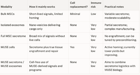
The full MSC secretome is likely to become the most comprehensive cell‑free platform because it captures exosomes, growth factors, cytokines, and non‑coding RNAs in a single product. Its multi-dose yield from a single harvest, long-term storage, preconditioning capability, and lack of live-cell risks make it highly attractive compared with both direct stem cell transplantation and exosomes alone.
The MUSE secretome adds another layer. Its unique patterns and exclusive proteins, including PZP and multiple stress-response proteins, suggest it is not merely a slightly better MSC secretome but a qualitatively different tool. Combined with MUSE cells’ ability to home to sites of damage and replace lost cells, the MUSE platform currently sits at the top of the regenerative hierarchy.
From a practical standpoint, the main hurdles now are not scientific but operational and regulatory: establishing consistent manufacturing standards, reliable potency assays, and clear approval pathways for secretome- and MUSE-based therapies. Clinically, a tiered approach makes sense: use isolated exosomes when their nanovesicle properties are essential, the full secretome when broad signaling is the goal, and the MUSE secretome or MUSE cells when durable engraftment and true structural repair are needed. Dr. Purita